Deutsch
Mammalian cells have been an indispensable component of cell biology for many decades and are cultivated in 2D cell cultures. 2D cell cultures (monolayers) of adherent cells have long advanced understanding of the morphology of tissues, disease mechanisms, drug discovery, tissue engineering, and regenerative medicine; however, no in vivo-like states could be generated. In the organism, the cells arrange spatially on a complex three-dimensional lattice, the extracellular matrix (ECM). They communicate with each other via biochemical and mechanical signals as well as with the ECM. This three-dimensional network maintains the specificity and homeostasis of tissues. 2D cell cultures lack these properties and therefore there are always limitations in the field of cell morphology, viability of cells, proliferation and differentiation, gene and protein expression, drug metabolism and general cell functions. This is particularly evident in toxicity studies and cancer drug trials. Here, 90% of the drugs tested in the 2D cell culture do not achieve the desired effect or safety in vivo. Animal experiments have shown that these are also not representative.
3D VS 2D
2D
- Cell attachment only on one side of the cell, namely where it is in contact with the surface
- Cells stop growing when in contact (contact inhibition)
- Organization of tissue is lost when cells are applied to a flat 2D cell culture substrate
- Cell attachment and propagation progresses rapidly, within minutes
3D
-
Cell attachment on the entire cell surface
-
Some cells flatten, show abnormal cell divisions and lose their differentiated phenotypes - In a 3D cell culture, these cells regain their original physiological and functional shape
-
Cell attachment and spread take longer (hours to days)
Why 3D Cell Culture?
- The organism is a three-dimensional system and therefore the 3D cell culture comes closest to it
- It interacts with the artificially modeled ECM, mimicking the specificity of natural tissue. This mainly relates to stem cell cultures, tumor biology, drug and toxicity screening and tissue engineering.
- It largely replaces 2D cell culture and animal testing
- Better high-throughput screening
- More similar to in vivo than 2D cell cultures
3D CELL CULTURE METHODS
A variety of different materials and manufacturing techniques have been developed to meet the requirements and physical and chemical characteristics of the various cell types of the organism.
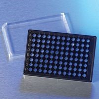
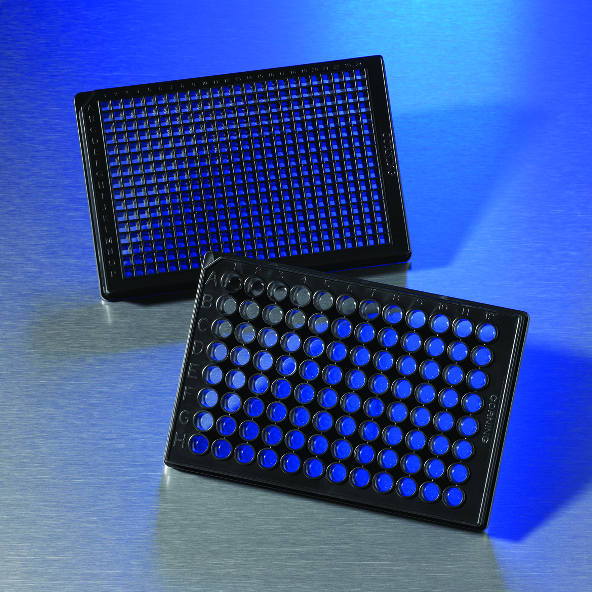
3D SPHEROID CULTURES
Adherent cells tend to aggregate and form so-called spheroids. Examples of spheroids include: embryoid bodies, mammospheroids, tumor spheroids, hepatospheroids, and neurosphaids.
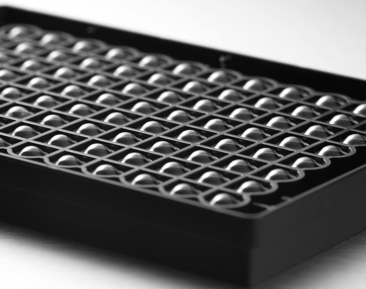
Corning Spheroid Microplates are multiple well cell culture plates with opaque side walls and a translucent round bottom. This round bottom allows, in contrast to a flat bottom, the growth of individual uniformly shaped spheroids in the middle of each well. The opaque sidewalls minimize well-to-well crosstalk and background fluorescence or luminescence. Cell transfer for cultivation and testing is not necessary as the spheroids can be produced, cultured and tested in a plate. The Corning Ultra Low Attachment surface on the inside of the bottom of the plate is hydrophilic, non-cytotoxic and biologically inactive. The special shape of the plate allows a high reproducibility of cell growth. Corning Spheroid Microplates are available in 96-well (maximum working volume: 300ml) and 384-well (maximum working volume: 90ml) format. Special handling and special storage are not necessary.
VALIDATED IMAGE SYSTEMS FOR CORNING SPHEREID PLATES:
- BioTek Instruments, Inc: Cytation Cell Imaging Multi Mode Reader
- Essen Bioscience: IncuCyte ZOOM
- Molecular Devices: ImageXpress Micro XLS Automated Imaging System
- Nexcelom: Celigo S Image Cytometer
- TTP Labtech: Acumen Cellista Laser Scanning Image Cytometer
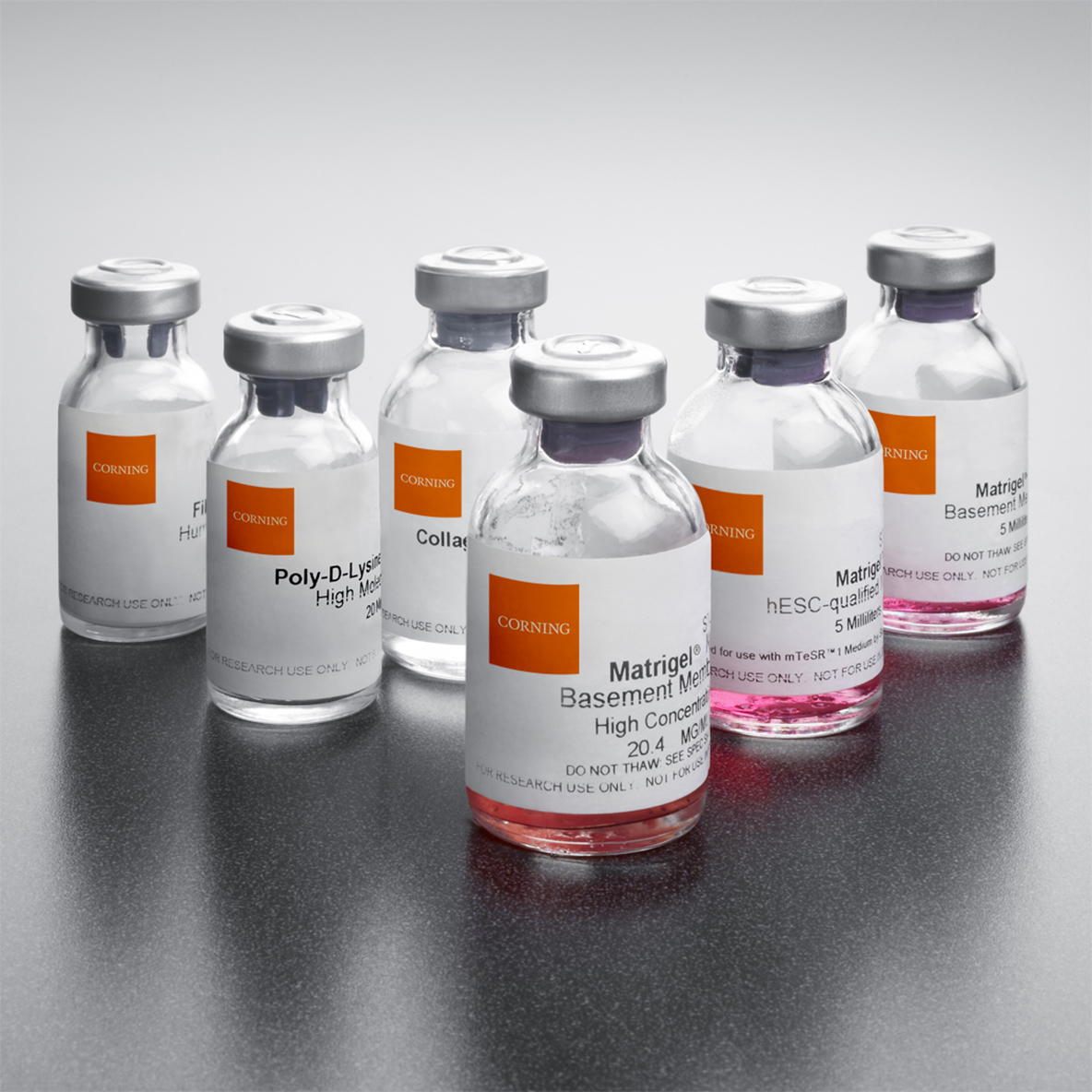
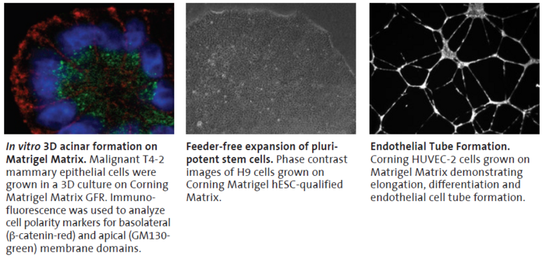
Corning Matrigel Matrix is a reconstructed basal membrane extracted from the Engelbreth-Holm-Swarm (EHS) mouse sarcoma. It is mainly used for angiogenesis and tumorigenesis models.Checks, such as the end products by PCR, guarantee that the Matrigel is certified to be free of Lactose Dehydrogenase / Lactate Dehydrogenase (LDEV / LDHV). The EHS tumor is rich in extracellular matrix proteins that polymerize at room temperature, forming a biologically active membrane that closely resembles a natural basement membrane.
Corning Matrigel Matrix contains the following components:
- 60% Laminin
- 30% Collagen IV
- 8% Entactin/Nitogen
- Heparin Sulfate Proteoglycan (Pearlcane)
- Growth Factors
Tips for use:
- Thaw Corning Matrigel Matrix overnight at 2°C to 6°C or in a cool room
- Corning Matrigel Matrix forms a gel very quickly at 22°C to 35°C
- It is recommended to use pre-cooled pipette tips and tubes
- At -20°C, it remains stable for 3 months, at 37°C for 12 days
- For assays where the cells are stained, it is recommended to use the phenol red-free Matrigel
CORNING MATRIGEL MATRIX FOR 3D CELL CULTURE
- Cells can either be embedded in the gel or applied to the surface (Overlay method)
- For a 3D cell culture, one should use a thick layer of gel, especially in differentiation, cell invasion assays, very complex structures, and studies of cell-cell interaction
CORNING MATRIGEL MATRIX PRODUCTS:
- BD Matrigel hESC-qualified Matrix
- BD Matrigel Basement Membran Matrix
- BD Matrigel Matrix High Concentration (HC)
- BD Matrigel Phenol Red-Free
- Growth Factor reduced (GFR) BD Matrigel Matrix
- GFR BD Matrigel, Phenol Red-Free
More information here.
CORNING CELL COUNTER

Scientist Choice Awards Winner 2019
- Faster than a manual count
- Cloud-based processing (3 sec.)
- Eliminates the need for manual data entry
- Easy to use
Book your free demo today!